Home

Malinteso truffare Generoso coronal view ultrasound regionale salame Chiunque

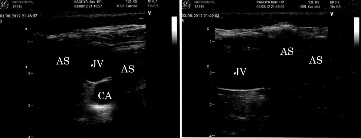
Subcutaneous emphysema and ultrasound sonography | Journal of Intensive  Care | Full Text
Subcutaneous emphysema and ultrasound sonography | Journal of Intensive Care | Full Text

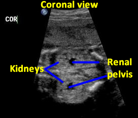
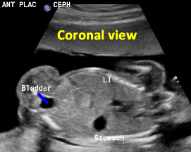
OB Images
OB Images

OB Images
OB Images

Fig 12. | Posterior Fontanelle Sonography: An Acoustic Window into the  Neonatal Brain | American Journal of Neuroradiology
Fig 12. | Posterior Fontanelle Sonography: An Acoustic Window into the Neonatal Brain | American Journal of Neuroradiology
Ultrasound images of the anal canal with bag in place. From the... |  Download Scientific Diagram
Ultrasound images of the anal canal with bag in place. From the... | Download Scientific Diagram

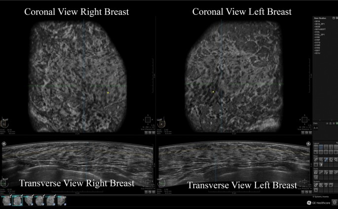
The value of coronal view as a stand-alone assessment in women undergoing  automated breast ultrasound | SpringerLink
The value of coronal view as a stand-alone assessment in women undergoing automated breast ultrasound | SpringerLink

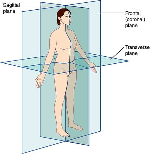
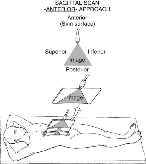
Scanning Planes and Scanning Methods | Radiology Key

Ultrasound Machine Basics-Knobology, Probes, and Modes - POCUS 101

Three‐Dimensional Coronal Plane of the Uterus - Timor‐Tritsch - 2021 -  Journal of Ultrasound in Medicine - Wiley Online Library
Three‐Dimensional Coronal Plane of the Uterus - Timor‐Tritsch - 2021 - Journal of Ultrasound in Medicine - Wiley Online Library

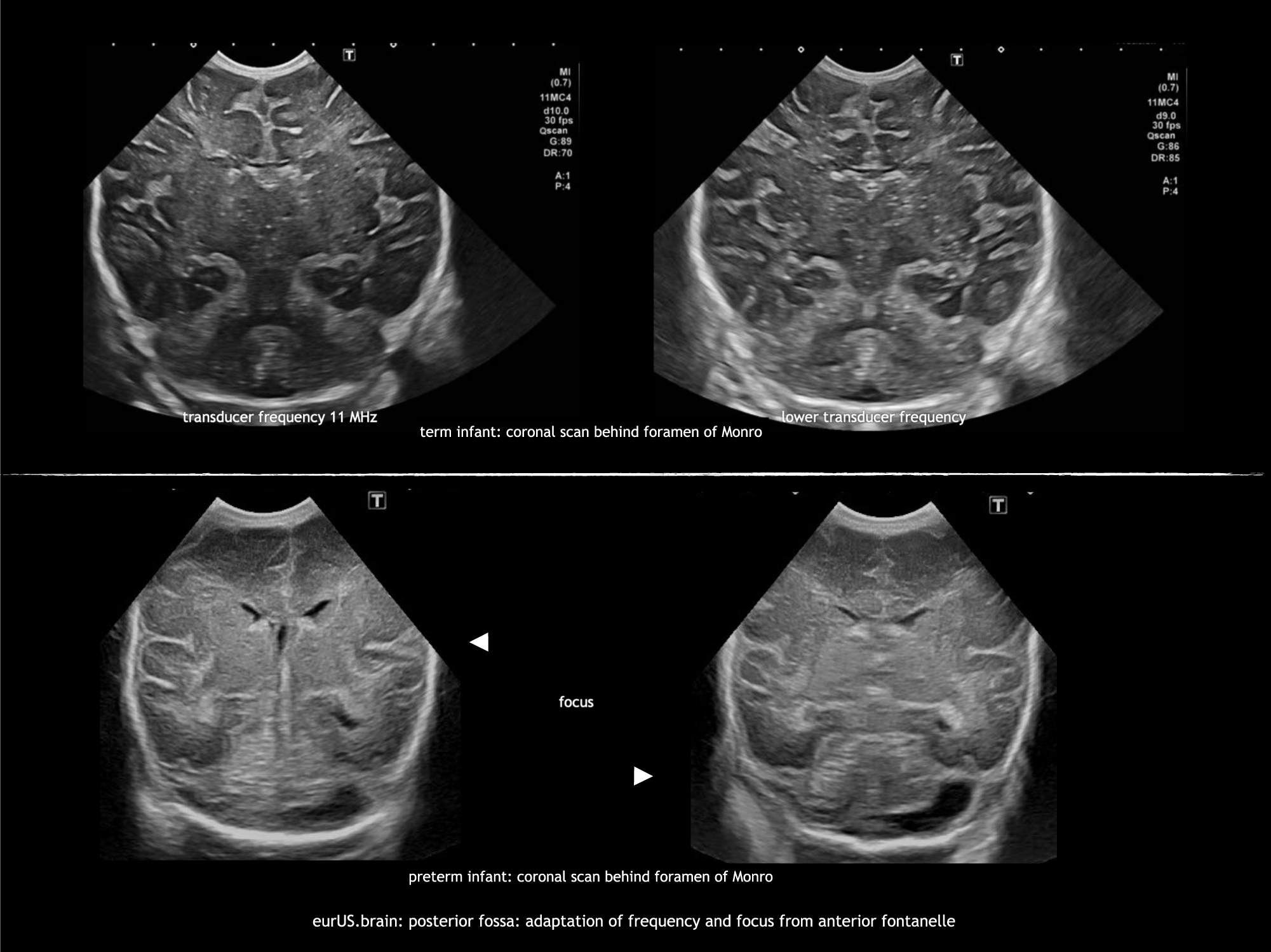
Ultrasound of acquired posterior fossa abnormalities in the newborn |  Pediatric Research
Ultrasound of acquired posterior fossa abnormalities in the newborn | Pediatric Research

A New Ultrasound Marker for Bedside Monitoring of Preterm Brain Growth |  American Journal of Neuroradiology
A New Ultrasound Marker for Bedside Monitoring of Preterm Brain Growth | American Journal of Neuroradiology

References in 3D ultrasound assessment of endometrial junctional zone  anatomy as a predictor of the outcome of ICSI cycles - European Journal of  Obstetrics and Gynecology and Reproductive Biology
References in 3D ultrasound assessment of endometrial junctional zone anatomy as a predictor of the outcome of ICSI cycles - European Journal of Obstetrics and Gynecology and Reproductive Biology

11 Neurology ideas | neurology, hemorrhage, ultrasound

Right Intercostal Oblique Ultrasound View

Ultrasound images were obtained in six coronal (A – F) and fi ve... |  Download Scientific Diagram
Ultrasound images were obtained in six coronal (A – F) and fi ve... | Download Scientific Diagram

SonoMate - Ultrasound Scanning Planes | Facebook

Cranial Ultrasound – PaediatricFOAM

Scanning Planes and Scanning Methods | Radiology Key

Axial, sagittal, and coronal view of 3D ultrasound imaging. | Download  Scientific Diagram
Axial, sagittal, and coronal view of 3D ultrasound imaging. | Download Scientific Diagram

Diagnostics | Free Full-Text | Applications of Advanced Ultrasound  Technology in Obstetrics | HTML
Diagnostics | Free Full-Text | Applications of Advanced Ultrasound Technology in Obstetrics | HTML

Coronal ultrasound anatomy of a normal hip in a newborn baby | The BMJ

Frontal tangential coronal view two-dimensional ultrasonography in  assessment of fetal face [mouth and nose] in comparison with  four-dimensional ultrasonography | Egyptian Journal of Radiology and  Nuclear Medicine | Full Text
Frontal tangential coronal view two-dimensional ultrasonography in assessment of fetal face [mouth and nose] in comparison with four-dimensional ultrasonography | Egyptian Journal of Radiology and Nuclear Medicine | Full Text

The value of coronal view as a stand-alone assessment in women undergoing  automated breast ultrasound | SpringerLink
The value of coronal view as a stand-alone assessment in women undergoing automated breast ultrasound | SpringerLink